Helen Maynard-Casely
The crystal structure rainbow – Green fluorescent protein
What does it look like?
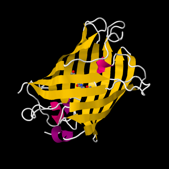
The GFP protein, drawn with Jmol. The protein folds into a cylindrical can (yellow) that houses the chromophore (shown as little balls) that results in the 'green' colouring of the protein when it is exposed to UV light.
What is it?
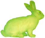
Alba the rabbit, whose genes were altered with GFP as part of work by artist Eduardo Kac.
In the waters of the North Pacific there is a group of jellyfish that have been floating about the ocean and gently glowing green. This remarkable feature is due to a particular protein, which was first extracted from the jellyfish Aequorea victoria in 1961. This work on Green Fluorescent Proteins (GFP) earned the scientists working on it a Nobel Prize for chemistry in 2008. The success of this work has been due to the incredible use that theses proteins have as markers in biochemistry. GFPs can be attached to viruses, for instance, and then – when illuminated by UV light – you can watch how the virus goes about its business and then design ways to halt its progress.
Where did the structure come from?
The GFP structure was first described by Ormö et al. in 1996, and can be found in the PDB, number 1EMA. Their work provided unparalleled insight into the inner workings of the protein. Since then researchers have investigated how to change the properties of GFP for particular applications. For example, engineered GFPs are now able to act as sensors to their surrounds and glow in response to differing environments, such as a change in acidity.
You can read lots more about GFP at the 'GFP website' or get more on their structure through PDB 101 article on them here.
Related articles
|
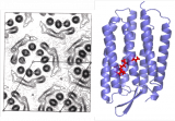
Protein crystals naturally occurring in a cellular membrane: Bacteriorhodopsin |
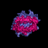
HLA Molecules – How the body detects infected cells |
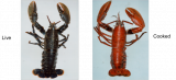
Why does a lobster change colour on cooking? |