Helen Maynard-Casely
Biominerals #2 – Weddellite (in Antarctica and in you!)
What does it look like?
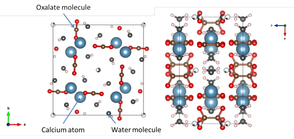
Two views of the Weddellite structure. Image generated by the VESTA (Visualisation for Electronic and STructural analysis) software http://jp-minerals.org/vesta/en/
What is it?
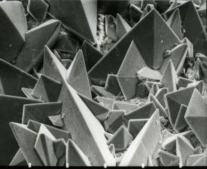
Scanning Electron Micrograph of the surface of a kidney stone showing tetragonal crystals of Weddellite. Image by Kempf EK.
This is another biomaterial that our own bodies form, and it can have some very painful consequences. Calcium oxalate is one of the materials that forms kidney stones, painful objects that can obstruct some of our internal organs. There are thought to be many reasons why these form, but the calcium oxalate is one of the minerals that conglomerates to form them.
There are a few ways that calcium atoms and oxalate molecules can crystallise together, and most of these involve water. The form we've featured today is the dihydrate. There are two water molecules in the structure for every calcium oxalate. This material actually has a mineral name, Weddellite, because as well as being found in your body it forms in sediment at the bottom of the Weddell sea in Antarctica.
Where did the structure come from?
This structure of Weddellite was determined in a paper that re-examined the crystal structure of weddellite and whewellite (the monohydrate (or one water atom per calcium oxalate) structure). It is #9000764 in the Crystallography Open Database.
Related articles
|
Classical crystal structures – sphalerite |
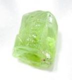
What is our planet made out of? (2) Olivine |
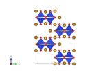
What is our planet made out of? (3) Wadsleyite |