Biological structures
Posted on 02/01/2014

Julia Archbold
HLA Molecules – How the body detects infected cells
What does it look like?
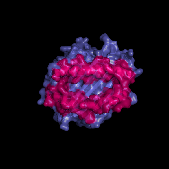
The structure of the class I human leukocyte antigen (HLA-A2) (pdb code: 3HLA http://www.rcsb.org/pdb/explore.do?structureId=3hla). Image generated by Pymol (http://www.pymol.org/ )
What is it?
During her PhD studies in Harvard, Pamela J. Björkman solved the structure of a protein (HLA) that tells the immune system whether a cell is healthy or infected. The studies by Björkman confirmed that both the protein AND the bound peptide are important for the recognition event, and supported the Nobel Prize winning work of Rolf Zinkernagel and Peter Doherty (see http://www.nobelprize.org/nobel_prizes/medicine/laureates/1996/)
The most beautiful part of this structure is the deep groove on its upper surface, encased by two alpha helical jaws (pink), which holds small peptides. The role of these HLA molecules is to present these peptides as sort of a barcode to be scanned by cells of the immune system. These peptides tell the immune system whether a cell is healthy and can be ignored, or infected and should be destroyed.
Where did the structure come from?
The structure was published in a 1987 Nature paper. It took Pamela eight trips to the German Synchrotron to solve the structure.
Related articles
|
The structure that started it all – Rock salt |
Tales from a PhD – Synthesising a catenane |
A protein structure that is a β-barrel of laughs |