Róisín McMahon
Lysozyme: Not to be sniffed at
What does it look like?
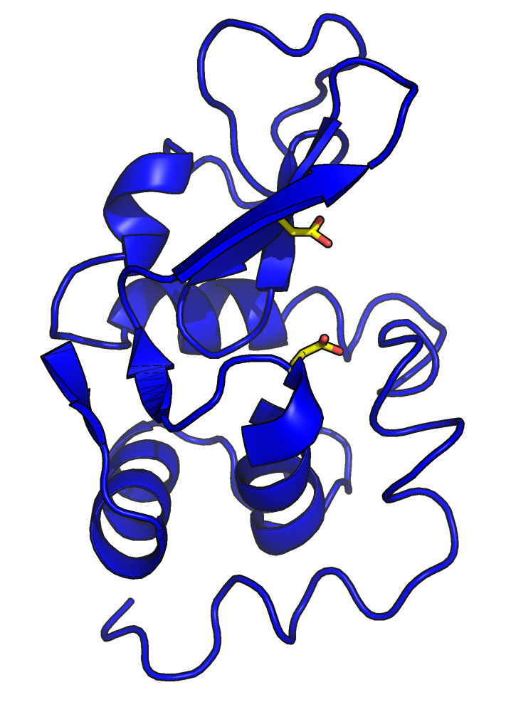
Cartoon representation of lysozyme from hen egg white (PDB ID 1LZY). The side chain atoms of two amino acids (glutamic acid 35 and aspartic acid 52) that are critical to the activity of the enzyme are show in yellow. The image was generated using the molecular graphics software PyMOL
What is it?
In 1922 Alexander Fleming, investigating the nasal secretion of a patient with acute coryza (i.e. a very runny nose), noticed that when the mucus was dropped onto a plate of bacteria it was able to inhibit growth of the bacteria completely. Fleming subsequently went on to show that the anti-bacterial activity he had observed was due to the enzyme lysozyme, and that it was also present in other human secretions. We now know that lysozyme's anti-bacterial activity stems from its ability to break down the chemical linkages between sugars in the bacterial cell wall, and that lysozyme is part of our first line of defence against infection.
Later in 1965 lysozyme became the second protein structure (and the first enzyme) to be determined by X-ray crystallography. The structure enabled a detailed mechanism of action for the enzyme to be described, the first time it had been done so for any enzyme and heralding a new era of understanding protein function through its structure.
In the almost 50 years since, lysozyme and its crystallisation have been extensively characterized, earning lysozyme a special place in the modern crystallographer’s tool box; lysozyme crystals are routinely used as model crystals by scientists when calibrating X-ray instrumentation, developing new methods of data collection, investigating the effects of radiation damage and when teaching students. Recently lysozyme has added another string to its bow with a role as a protein-engineering tool; insertion of T4 lysozyme into the third intracellular loop of a G-protein coupled receptor (GPCR) assisted crystallisation of a member of this very important family of membrane proteins. This work has revolutionised the field of GPCR structural studies, and one of its pioneers is the 2012 Nobel Laureate for Chemistry, Brian Kobilka (who shared the prize with Robert Lefkowitz.)
All told, a nifty little trick to have up your nose.
Where did the structure come from?
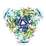
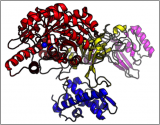
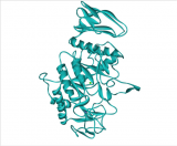
There are more than a thousand different crystal structures of lysozyme in the Protein Data Bank, made up of mutants and variants from different organisms including human, mouse and the bacteriophage T4. This structure is a refined model of the original structure solved in 1965 in which the protein was isolated from hen egg white, which is naturally abundant in lysozyme.
Related articles
|
Crystallising enzymes from beans – celebrating Sumner |
From crystal to structure in 84 years |
The Start of Digestion: Salivary α-Amylase |