Katy Wood
Protein crystals naturally occurring in a cellular membrane: Bacteriorhodopsin
What does it look like?
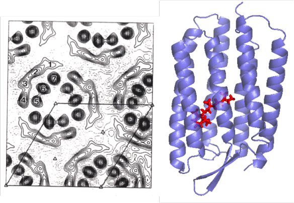
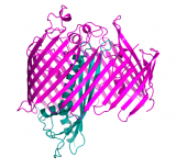
On the left: an electron micrograph of the naturally occurring two-dimensional crystal of the protein, taken directly from cellular membrane (Unwin & Henderson 1975). Dark 'blobs' are the protein (bacteriorhodopsin), in between are lipids. On the right is one of the more recent crystal structures of bactiorhodopsin (Belrhali et al. 1999), PDB entry 1QHJ (Figure made with Pymol). The 'blobs' in the micrograph are helices viewed from above – these helices can be seen in the transmembrane view in the structure on the right.
What is it?
Bacteriorodopsin is one of life's amazing little energy machines: a photosensitive molecule in the middle of the protein takes energy from the sun and uses it to pump protons across the cell membrane.
Membrane proteins are notoriously hard to study, but nature at least gave us one gift in this regard: a protein that naturally forms a two-dimensional crystal structure in a membrane – bacteriorhodopsin.
Because it could be purified directly in a crystalline form, bacteriorhodopsin was one of the first membrane proteins to be studied structurally in great detail by a variety of techniques including electron microscopy and neutron diffraction.
Many stages of the machine's 'pumping' action (intermediate structures) have now been resolved by X-ray crystallography, making this a great example of how structures don't just give us pretty pictures – they show us how life's little machines work.
Where did it come from?
Bacteriorhodopsin has been extensively studied by too many groups to cite, who all contributed to understanding its mechanics. The image of the 2-D crystal on the left is from one of the very early electron microscopy studies, the structure on the right is from 3-D crystals of the protein grown with lipids.
References:
Henderson R, Unwin PN (1975) Three-dimensional model of purple membrane obtained by electron microscopy. Nature, 4; 257:28-32.
Belrhali H, Nollert P, Royant A, Menzel C, Rosenbusch JP, Landau EM, Pebay-Peyroula E (1999) Protein, lipid and water organization in bacteriorhodopsin crystals: a molecular view of the purple membrane at 1.9A resolution. Structure, 7, 909-917.
Related articles
|
'Roll out the Barrel': Structure of the LptD-LptE translocon complex from bacteria |
Cold as Ice… with 'Maxi' sacrifice |
Spinning around with Spinels – Lithium titanate |