Jonathan McCree-Grey
β-Carotene helps you see in the dark
What does it look like?
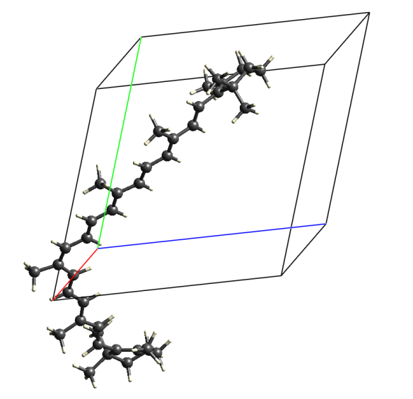
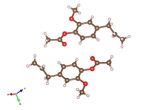
Image generated using Crystal Explorer 3.1 using data available from the Crystallography Open Database (#2016501). Crystals are of a monoclinic P21/c structure with two molecules in the unit cell.
What is it?

Beta-carotene is a red-orange pigment found in many plants and fruit and is what gives carrots, pumpkins and sweet potatoes their bright orange colour. It was originally named by H. Wachenrӧder in 1831, who was the first to isolate and crystallise the molecule from the roots of carrots, though it may also be recognisable as E-number E160a, which it is assigned when used as a food colouring.
β-carotene is a natural precursor for vitamin A, needed by the retina of the eye in the form of retinal, which combines with the protein opsin to produce rhodopsin, a light absorbing molecule necessary for low light and colour vision. However, you don't want to digest too much β-carotene, as overconsumption can cause Carotenosis, a benign condition which turns your skin orange!
Where did the structure come from?
Preliminary X-ray studies of β-carotene were conducted in 1937 and 1948; however, neither of these attempts were able to successfully resolve the system's molecular structure. It wasn’t until 1964 that that the actual atomic arrangement of beta-carotene was first determined (C. Sterling Acta. Cryst. 1964 17, 1224). The data for the structure presented here is from a much more recent study, from 2008, and is available from the Crystallography Open Database (# 2016501).
Related articles
|
Pinene – It's beginning to smell a lot like Christmas! |
Adapting a plant-based molecule – eugenyl acetate |
Five fold - Cobalt tricyanomethanide |