Anton Le Brun
A Crystal Structure for Stopping Germ Warfare
What does it look like?
Protective antigen from Bacillus anthracis.
What is it?
Anthrax is the disease that is caused by the soil bacteria Bacillus anthracis. Anthrax commonly infects wild and domestic animals such as cattle, which ingest bacterial spores whilst grazing, but anthrax can also (rarely) infect humans. B. anthracis ability to form spores to survive extreme environments is what makes it suited to turn it into a biological weapon. This is where anthrax gets its notoriety from. One example includes the 2001 Anthrax attacks where B. anthracis spores were posted in letters to various US news outlets and US Senators.
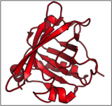
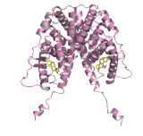
When the spores get inside the body they are activated. The bacteria start to grow and multiply and produce the anthrax toxin. There are three proteins that make up the anthrax toxin: protective antigen, edema factor and lethal factor. Today we are showing you the structure of protective antigen (so named because it is the antigen that is used in the current anthrax vaccine). Protective antigen is an 83 kDa protein that has four domains. Domain I (cyan) has a jelly roll topology, domain II (pink) has a Greek key topology, domain III (orange) has a similar topology to ferredoxin, and domain IV (green) has an immunoglobulin-like topology.
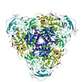
When protective antigen binds to receptors on the cell surface of the infected host the protective antigen is cleaved in domain I by a protein called furin, creating two fragments: PA63 and PA20. PA20 plays no further role in the anthrax story but PA63 will form a heptamer (a group of seven PA63 molecules, see below) which edema factor and lethal factor bind to. The PA63 heptamer with edema and lethal factors bound is internalised into the cell in an endosome. The low pH inside the endosome causes the PA63 heptamer to change in conformation and form a pore releasing edema factor and lethal factor into the cell. Edema factor disrupts water homeostasis causing a build-up of fluids in the body known as edema, and lethal factor disrupts cell signalling process which leads to cell death.
In 1881 Louis Pasteur demonstrated the use of the first vaccine against anthrax (and this was the second vaccine to be made after Edward Jenner's smallpox vaccine in 1796). Pasteur's vaccine was a weakened strain of B. anthracis by exposing the bacteria to oxygen (although there is some controversy surrounding this). Modern anthrax vaccines are a bit more sophisticated and protective antigen forms part of the active ingredient.
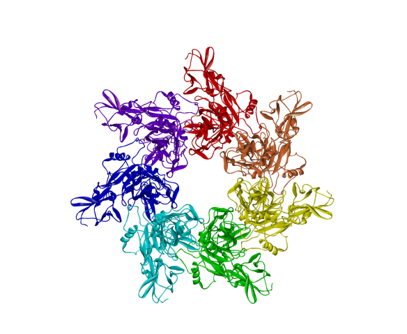
The B. anthracis PA63 heptameric pre-pore.
Where does it come from?
The crystal structure of protective antigen was first solved by Carlo Petosa and colleagues in 1997 [1] (PDB number 1ACC) and the crystal structure of the PA63 heptamer pre-pore was solved by Borden Lacy and colleagues in 2004 [2] (PDB number 1ZTO).
[1] C. Petosa, R.J. Collier, K.R. Klimpel, S.H. Leppla and R.C. Liddington (1997) Crystal structure of the anthrax toxin protective antigen. Nature 385: 833-838.
[2] D.B. Lacy, D.J. Wigelsworth, R.A. Melnyk, S.C. Harrison and R.J. Collier (2004) Structure of heptameric protective antigen bound to an anthrax toxin receptor: a role for receptor in pH-dependent pore formation. Proceedings of the National Academy of Sciences USA 101: 13147-13151.
Related articles
|
Crystallising enzymes from beans – celebrating Sumner |
Oh, deer... |
'Let's talk about Sex' hormones – Part 1: The Oestrogen Receptor |