Anton Le Brun
From Toxin to Technology: α-Haemolysin
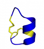
What does it look like?
α-Haemolysin from Staphylococcus aureus with each segment coloured a different colour of the rainbow.
What is it?
Today's post is α-haemolysin (or α-hemyolsin for our American readers); a toxin produced by the bacteria Staphylococcus aureus which is associated with many illnesses, the more serious including sepsis and pneumonia.
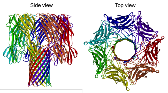
As the name suggests α-haemolysin attacks red blood cells. The α-haemolysin toxin is secreted by the S. aureus bacteria as monomers, and once bound to the red blood cell membrane will form a heptamer (a group of seven). The α-haemolysin heptamer forms a large mushroom shaped structure which forms a hole in the cell membrane. The contents of the cell leak through this hole, eventually killing the cell.
α-Haemolysin is a really good example of turning a protein found in nature into something technologically useful. Scientists have engineered α-haemolysin to form nanopores that can sequence DNA. The DNA strand is passed through the hole formed by haemolysin in the presence of an electrical current. When the DNA is passed through the pore this changes the electrical conductance and the four nucleotides that make up the DNA alphabet (A, T, C and G) each change the conductance differently allowing for the DNA to be sequenced [1].
Where does it come from?
The α-Heamolysin structure was solved by L. Song and colleagues in 1996 and has the PDB reference 7AHL [2].
[1] D. Branton et al. The potential and challenges of nanopore sequencing (2008) Nature Biotechnology 26: 1146 – 1153
[2] L. Song et al. The structure of Staphylococcal alpha-hemolysin, a heptameric transmembrane pore (1996) Science 274: 1859 -1866
Related articles
|
DNA: Life's blueprint |
From crystal to structure in 84 years |
Cone snail toxins as painkillers – The structure of an alpha-conopeptide |