John Helliwell
Celebrating Laue – comprehensive protein structures
In the last of our three posts celebrating Max von Laue and Laue diffraction, Prof. John Helliwell tells us how the technique has impacted on protein crystallography.
What is it?
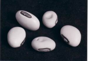
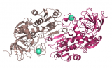
Concanavalin A is isolated from jack beans; approx 10% by weight of protein is con A. Its role is not known for sure but is implicated in an antifungal protection strategy for the bean via protein to protein cross linking involving polysaccharide.
To get a comprehensive picture of a protein structure in its functional state(s) can lead to seeking a time-resolved 'movie'. But the X-ray data collection may be too slow to capture the interesting moment of function. As an alternative to use of monochromatic radiation the use of polychromatic X-ray synchrotron radiation leads to much quicker diffraction data collection. Moreover the sought-after hydrogenation and bound water details may be incomplete as hydrogen scatters X-rays weakly. But this latter challenge can be overcome by use of neutron diffraction, as deuterium scatters neutrons as strongly as e.g. carbon. But neutron beams are much weaker in intensity than synchrotron X-ray beams.
As in the X-ray case above, though, the polychromatic neutron emission from either a nuclear reactor or a pulsed source can be more efficiently harnessed using Laue diffraction and again leads to shorter exposure times. Research in the 1980s [1,2] led to a much improved understanding of Laue crystallography and greatly assisted accurate Laue diffraction data analysis along with newly developed software.
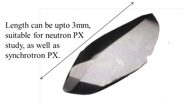
Overall this has led to a variety of time-resolved Laue X-ray protein crystallography and neutron Laue crystallography studies [e.g. 3,4]. As just one example the crystal shown here, ~3mm long, is of the lectin protein concanavalin A (isolated from jack beans ~1 cm, also pictured), and was used both to help develop these modern Laue methods and to reveal new details of its bound water structure at the sugar molecular recognition binding site both at room temperature and at 15K [4].
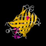
What does the structure look like?
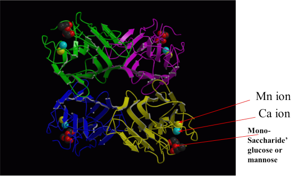
In a final twist of this story the neutron Laue study of concanavalin A at 15K [4] showed that freeze quench time-resolved studies with neutrons even of large crystals of a protein was possible. In the ten years since this study [4] there have been various further improvements in neutron beam fluxes, measuring apparatus and use of fully deuterated proteins in Europe, Japan and the USA, radically changing the scope of neutron macromolecular crystallography to smaller crystals and larger molecular weights of proteins.
[1] D.W.J. Cruickshank, J.R. Helliwell and K. Moffat 'Multiplicity Distribution of Reflections in Laue Diffraction'. Acta Cryst. (1987), A43, 656-674.
[2] D.W.J. Cruickshank, J.R. Helliwell and K. Moffat 'Angular distribution of reflections in Laue diffraction' Acta Cryst. (1991), A47, 352–373.
[3] J.R. Helliwell, Y.P. Nieh, J. Raftery, A. Cassetta, J. Habash, P.D. Carr, T. Ursby, M. Wulff, A.W. Thompson, A.C. Niemann and A. Hädener “Time–resolved structures of hydroxymethylbilane synthase (Lys59Gln mutant) as it is loaded with substrate in the crystal determined by Laue diffraction” (1998) Faraday Trans. 94(17), 2615–2622.
[4] M.P. Blakeley, A.J. Kalb (Gilboa), J.R. Helliwell & D.A.A. Myles (2004) “The 15-K neutron structure of saccharide free concanavalin A” Proceedings of the National Academy of Sciences USA 101, 16405-16410. PDB code 1XQN.
Related articles
|
The crystal structure rainbow – Green fluorescent protein |
Bak: The Face of Death |
Breaking down the drink – Alcohol Dehydrogenase |